
Iñigo Gabilondo, Verónica Llorens, Trinidad Rodriguez, Manuel Fernández, Tomas Pérez Concha, Marian Acera, Beatriz Tijero, Ane Murueta-Goyena, Rocío del Pino, Jesús Cortés, Juan Carlos Gómez-Esteban. Myocardial MIBG scintigraphy in genetic Parkinson’s disease as a model for Lewy body disorders. European Journal of Nuclear Medicine and Molecular Imaging, 46:376-384, 2019 [pdf]
Purpose. To identify myocardial sympathetic denervation patterns suggestive of Lewy body (LB) pathology in patients with genetic and idiopathic parkinsonisms by 123I-metaiodobenzylguanidine (MIBG) scintigraphy.
Methods. We retrospectively analysed myocardial MIBG images acquired with a dual-head gamma camera and low-energy high- resolution collimator (LEHR) in 194 patients with suspected synucleinopathy or atypical parkinsonism, including 34 with genetic Parkinson’s disease (PD; 4 PARK1, 8 PARK2 and 22 PARK8), 85 with idiopathic PD (iPD), 6 with idiopathic REM sleep behaviour disorder (iRBD), 17 with dementia with LB (DLB), 40 with multiple system atrophy (MSA) and 12 with progressive supranuclear palsy (PSP), and in 45 healthy controls. We calculated heart-to-mediastinum MIBG uptake ratios (HMR) at 15 min and 4 h (HMR4H) for the LEHR and standardized medium-energy collimators, to obtain classification accuracies and optimal cut-off values for HMR using supervised classification and ROC analyses.
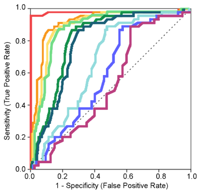
Results. While patients with LB disorders had markedly lower HMR4HLEHR than controls (controls 1.86 ± 0.26, iPD 1.38 ± 0.29, iRBD 1.23 ± 0.09, PARK1 1.20 ± 0.09, DLB 1.17 ± 0.11; p < 0.05), for the remaining patient categories differences were smaller (PARK8 1.51 ± 0.32; p < 0.05) or not significant (MSA 1.82 ± 0.37, PSP 1.59 ± 0.23, PARK2 1.51 ± 0.30; p > 0.05). The diag- nostic accuracy of HMR4HLEHR was highest in patients with LB disorders (PARK1, iPD, DLB, iRBD; 89% to 97%) and lowest in those with PARK2, PARK8, PSP and MSA (65% to 76%), with an optimal HMR4HLEHR cut-off value of 1.72 for discrim- inating most patients with LB disorders including iPD and 1.40 for discriminating those with aggressive LB spectrum phenotypes (DLB, PARK1 and iRBD).
Conclusion. Our study including patients with a wide spectrum of genetic and idiopathic parkinsonisms with different degrees of LB pathology further supports myocardial MIBG scintigraphy as an accurate tool for discriminating patients with LB spectrum disorders.