
Javier Rasero, Hannelore Aerts, Marlis Ontivero Ortega, Jesus M. Cortes, Sebastiano Stramaglia, Daniele Marinazzo. Functional Network prediction from region connectivity profiles and application to brain tumor data. OHBM 2019 – Organization for Human Brain Mapping [pdf]
Introduction:
Intrinsic Connectivity Networks (ICNs), patterns of correlated activity emerging from “resting- state” BOLD time series, are increasingly being associated with cognitive, clinical, and behavioral aspects, and compared with the pattern of activity elicited by specific tasks. ICNs examples include the visual and motor system and the default mode network. The ability to reliably estimate these functional networks from resting-state fMRI data would have important clinical implications, for example in neurosurgeons’ pre-surgical planning when patients are unable to cooperate with the task-based paradigm. As a consequence, adopting a supervised machine learning approach to select the most optimal model, we attempt to quantify to which extent one can predict ICNs training in task-based paradigm and predict the ICN patterns emerging purely from resting-state fMRI data. The development of a general framework for ICN identification could thus shed further light on the overlap between the patterns emerging from both task and resting-state fMRI data and elucidate topological cognitive changes due to brain lesions.
Methods:
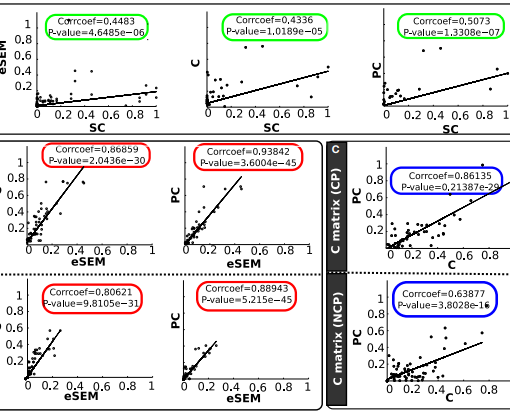
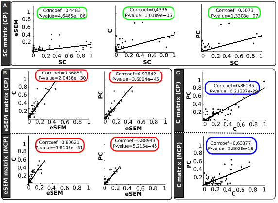
We computed resting-state and task-based fMRI correlation maps from 282 unrelated healthy subjects provided by the Human Connectome Project. Each of these maps, whose label is known, is composed of 268 features and was used to identity ICNs by means of four well-known machine learning algorithms. Out-of-sample performance was estimated on resting fMRI data, acting thus as the unseen test set, with the aim of addressing the question of how accurate these predictions are when they are obtained from a classifier fixed on task fMRI, acting here as the training set. Likewise, this latter dataset was previously used to vary the model hyperparameters and select the best classifier in a 5-times repeated 10-fold cross-validation, each hold-out pieces of data in each fold acting thus as the validation set. Likewise, the best model was later used to address topological changes in ICN assignment in 22 subjects with brain tumor data.
Results:
First, a neural network with four hidden layers of 512, 256, 128 and 64 units provides the best performance in the model selection step on task fMRI correlation maps. All the ICNs exhibit a good accuracy, especially those networks whose activation is usually demanded during our task performance, achieving all rates above 90%, except for the limbic system in which the accuracy drops to approximately 66%, with a remarkable 18% of examples misclassified as subcortical regions. Second, this optimal and already trained model generalises well in resting data where no active collaboration of the subjects is required, exhibiting a similar performance, with both Visual and Sensorimotor networks reaching prediction rates of approximately 92% and 91% of sensitivity respectively. Similarly, attention areas exhibit a 84% and 79% performance respectively, whereas DMN regions whose activity negatively correlates with that of the other networks during rest also exhibit a decent performance of about 79%, which suggests that these areas maintain an intrinsic correlation that allows them to be optmially fitted by a model even during task. Finally, this methodology also reveals important ICN label changes in brain regions due to brain tumor development.


