
Garazi Labayru, Ibai Diez, Jorge Sepulcre, Esther Fernández, Miren Zulaica, Jesús M. Cortés, Adolfo López de Munain, Andone Sistiaga. Regional brain atrophy in gray and white matter is associated with cognitiveimpairment in Myotonic Dystrophy type 1 Neuroimage: Clinical 24: 102078, 2019 [pdf]
Abstract
Background
Myotonic Dystrophy type 1 (DM1) is a slowly progressive myopathy characterized by varying multisystemic involvement. Several cerebral features such as brain atrophy, ventricular enlargement, and white matter lesions (WMLs) have frequently been described. The aim of this study is to investigate the structural organization of the brain that defines the disease through multimodal imaging analysis, and to analyze the relation between structural cerebral changes and DM1 clinical and neuropsychological profiles.
Method
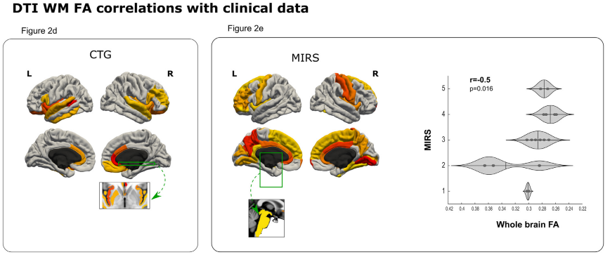
31 DM1 patients and 57 healthy controls underwent an MRI scan protocol, including T1, T2 and DTI. Global gray matter (GM), global white matter (WM), and voxel-level Voxel Based Morphometry (VBM) and voxel-level microstructural WM abnormalities through Diffusion Tensor Imaging (DTI) were assessed through group comparisons and linear regression analysis with age, degree of muscular impairment (MIRS score), CTG expansion size and neuropsychological outcomes from a comprehensive assessment.
Results
Compared with healthy controls, DM1 patients showed a reduction in both global GM and WM volume; and further regional GM decrease in specific primary sensory, multi-sensory and association cortical regions. Fractional anisotropy (FA) was reduced in both total brain and regional analysis, being most marked in frontal, paralimbic, temporal cortex, and subcortical regions. Higher ratings on muscular impairment and longer CTG expansion sizes predicted a greater volume decrease in GM and lower FA values. Age predicted global GM reduction, specifically in parietal regions. At the cognitive level, the DM1 group showed significant negative correlations between IQ estimate, visuoconstructive and executive neuropsychological scores and both global and regional volume decrease, mainly distributed in the frontal, parietal and subcortical regions.
Conclusions
In this study, we describe the structural brain signatures that delineate the involvement of the CNS in DM1. We show that specific sensory and multi-sensory — as well as frontal cortical areas — display potential vulnerability associated with the hypothesized neurodegenerative nature of DM1 brain abnormalities.
