
Ane Murueta-Goyena , Roco del Pino, Paula Reyero , Marta Galds, Begoa Arana, Olaia Lucas Jimenez, Marian Acera, Beatriz Tijero, Naroa Ibarretxe-Bilbao, Natalia Ojeda, Javier Pea, Jesus Cortes, Juan Carlos Gomez-Esteban, Iñigo Gabilondo. Parafoveal thinning of inner retina is associated with visual dysfunction in Lewy body diseases. Movement disorders 34:1315-1324, 2019 [pdf]
Abstract
Background: Retinal optical coherence tomography findings in Lewy body diseases and their implications for visual outcomes remain controversial. We investigated whether region-specific thickness analysis of retinal layers could improve the detection of macular atrophy and unravel its association with visual disability in Parkinson’s disease.
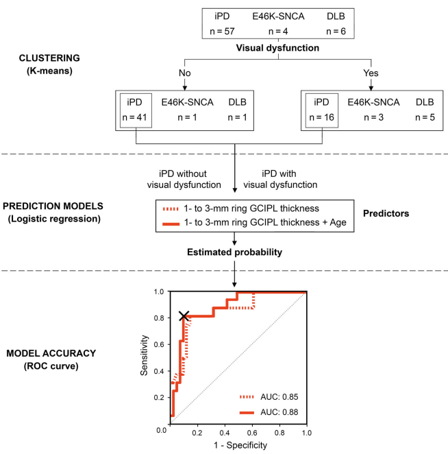
Methods: Patients with idiopathic Parkinson’s disease (n = 63), dementia with Lewy bodies (n = 8), and E46K mutation carriers in the α-synuclein gene (E46K-SNCA) (n = 4) and 34 controls underwent Spectralis optical coherence tomography macular scans and a comprehensive battery of visual function and cognition tests. We computed mean retinal layer thicknesses of both eyes within 1-, 2-, 3-, and 6-mm diameter macular discs and in concentric parafoveal (1- to 2-mm, 2- to 3-mm, 1- to 3-mm) and perifoveal (3- to 6-mm) rings. Group differences in imaging parameters and their relationship with visual outcomes were analyzed. A multivariate logistic model was developed to predict visual impairment from optical coherence tomography measurements in Parkinson’s disease, and cutoff values were determined with receiver operating characteristic analysis.
Results: When compared with controls, patients with dementia with Lewy bodies had significant thinning of the ganglion cell-inner plexiform layer complex within the central 3-mm disc mainly because of differences in 1- to 3-mm parafoveal thickness. This parameter was strongly correlated in patients, but not in controls, with low contrast visual acuity and visual cognition outcomes (P < .05, False Discovery Rate), achieving 88% of accuracy in predicting visual impairment in Parkinson’s disease.
Conclusion: Our findings support that parafoveal thinning of ganglion cell-inner plexiform complex is a sensitive and clinically relevant imaging biomarker for Lewy body diseases, specifically for Parkinson’s disease. © 2019 The Authors. Movement Disorders published by Wiley Periodicals, Inc. on behalf of International Parkinson and Movement Disorder Society.